 |
| Happy days: PhDing at the NHM with the holotype of Polacanthus foxii. |
We rightly hear a lot about early career researchers (ECRs) and the trials they face as they embark on their careers as scientists and academics, a daunting and challenging time in any scientist’s life. However, there is also a smaller group of people active in academia who are a bit longer in the tooth and might well have already had a career, or at least still be in the middle of one. These are the mature students, and I was one of them.
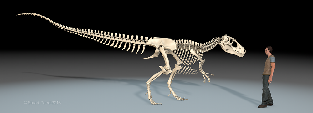
I came to academia late in life. I was in my early 40’s when I realised I wanted to become engaged in paleontological research in a more formal and structured manner than just picking bits of dinosaur bone off the beach on my frequent trips to the Isle of Wight (an activity I still enjoy). In 2010 I went on a week’s dig on the Hell Creek, followed a year later by the SVP at Las Vegas and from there became a research associate at the National Oceanography Centre Southampton (NOCS), part of the University of Southampton. I had tried to apply for an MRes course but it was suggested I didn’t have the qualifications. Actually, that’s correct. I have three ‘O’ levels, a general art and design certificate and a B/TEC in Graphic Design… and that’s the lot (I fluffed several ‘O’ levels because I was bad at exams and got in a panic; I’ve been an advocate of continuous assessment ever since). But I had relevant “life experience” after a long career in graphics. I was an active paleontological researcher and had presented my work and co-hosted workshops at international meetings etc
Growing up, university was never mentioned at school or home, except once when my physics teacher told me he’d buy me pint when I got my degree. University was not on the radar at all, it was an option so out of my reach that it was never discussed, ever. It never occurred to me I could go on to higher education. So I went to art school, got my graphic design qualification and that’s been my career for over 30 years, the last 14 of them running my own business.
When I was offered the opportunity to study for a self-funded, part-time vertebrate palaeontology PhD at NOCS, I was both surprised and excited. I went through the process of application, had my interview (toughest I’d ever done) and to my joy and amazement was accepted. I was very happy; here was a chance to make up for opportunities I didn’t have when younger. I was the first of my family to go to university.
Part of the reason I left salaried employment to run a business was that I wanted to study more, but the reality of being a sole trader made that difficult to achieve, and this became clear in the first couple of years. Structured courses were problematic as my day job comes first and I ended up missing exams etc to meet deadlines; weekends were spent working and lessons would be missed, and even part-time distance learning proved to much. Work comes first, end of. No work, no money.
The PhD was different. I could plan my research and even if I didn’t go near it for months could come back when things were quiet and resume without problem. Even on days I was stacked with work I could move my research on even if only incrementally. I’d send an email, download a paper, make a note of something, process data on an idle machine or any other of the myriad of activities a PhD student has do. It didn’t impact my work, but the tiny tasks returned tangible results, gave me a sense I was still progressing and during downtime I could make good headway. I enjoyed this period of my degree immensely. I was learning and becoming part of the wider community of palaeontologists who work on vertebrates. Even though I was 200 miles from the department I felt part of the Vertebrate Palaeontology (VP) group. The PhD began to feed back into my day job in ways I'd never anticipated, but were enormously beneficial.
Our VP group were a busy bunch. We hosted international vert palaeo meetings, we talked about our research, presented posters, taught and gave workshops and I co-supervised a brilliant MRes student. I was making good progress, my panel were happy and I was preparing to upgrade, and then my supervisor left, suddenly and without warning. Following this event issues with the specimen I was working on came to light (and was totally unaware of when I started), and over a period of several months my PhD imploded completely.
If this wasn’t bad enough, the university really didn’t care. Understandably and rightly it was the ECRs who needed to be supported most; their future careers depended on getting through the months that followed our supervisor’s departure. This was the priority, and I’m pleased to say they have all graduated. Liz Martin Silverstone, another member of the team, posted her thoughts on what could be learnt from her experience in this excellent blog post: https://gimpasaura.wordpress.com/2018/01/02/things-i-learned-during-my-phd-dos-and-donts-for-students-to-staff/
Things went from bad to worse. The senior staff simply did not seem to give a damn about my PhD. In my first call to one of them when I told her I had no undergrad or masters her exact words were; “How did you get in?”. It was suggested I could move to an MPhil instead as a consolation; my PhD was dead in the water, they had decided. The issues with the specimen meant that even this became impossible and the legal department pretty much stopped answering my emails. I became stressed, developed alopecia (my beard hair fell out in patches; it’s grown back now) and in the face of utter indifference from the senior staff I withdrew; I had no choice. My fees were returned but it was a financial disaster as I’d spent as much again on my doctorate, an investment with no return. As for the time invested… best not think about that.
I probably could have handled it better in some ways. I am very honest with my clients, and this sort of candour which is essential in business was not appreciated by the senior staff. I reply to emails, I keep people informed and if there is an issue I let them know at the earliest possible opportunity, however difficult this might be to inform them of. I didn’t know how an institution like a university works as I’d not experienced it before. I was angry and upset and this hinders communication (I still am, and having spoken to friends whose PhDs were also ended through no fault of their own it’s likely I will be for a long time).
It’s an oft-repeated myth that for those of us without a degree that life experience is taken into account when older students apply for higher degrees. True, I DID get accepted at NOCS on this basis (my day job meant there was some crossover in both the technology and ability to plan and execute large projects), but had the person I spoke to on the phone been in charge, I’m inclined to think I wouldn’t have. I’m now not sure I ever will again.
Universities have a duty of care to all their students, from ECRs building careers in a competitive and challenging environment to the late comers who want to try to realise some of their own potential that they weren't in a position to earlier in life. Older students can find universities intimidating places (EVERYONE is so much cleverer than you) and when things go wrong senior staff have to treat mature students as equals and with respect, not as some sort of irritating distraction. I was deadly serious about my PhD, but it turns out the senior staff at the school were not. All students, including the older ones, put their trust in senior staff and do not expect to be let down in such a manner, and in the case of the VP group we all were.
Positives? There are plenty. I learned a lot and loved being a VP PhD student. Most other palaeontologists have been fantastically supportive and understanding of the situation and I am looking forward to the future with an exciting project in the pipeline, working with people that I admire and can learn from and I look forward to contributing to the science. For a non-academic academic, I’m lucky to have such an opportunity. More on that later.
Had I failed at my PhD because I couldn’t complete for some reason of my own, such as being intellectually or academically incapable, financially compromised or whatever else that would have been one thing, but to fail because of the (in)actions and indifference of those who are in a position to ensure all students are helped through tough times? That should worry all of us.